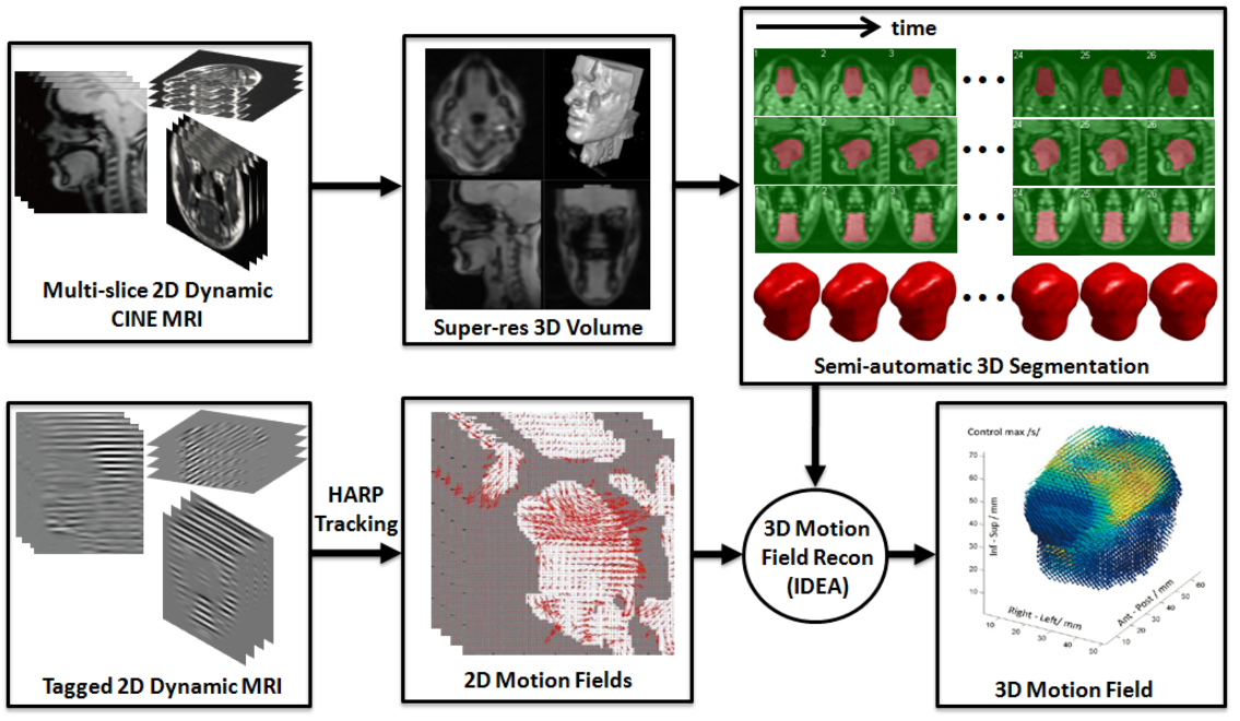
4D-MRI based Tongue Motion Analysis after Tongue Cancer Surgery Oral, head and neck cancer represents 3% of all cancers in the United States and is the 6th most common cancer worldwide. Recent studies have shown a five-fold increase in the incidence of squamous cell carcinomas of the oral portion of the tongue among young men and a six-fold increase among young women. Glossectomy is the surgical removal of a cancerous tumor. The amount of tongue tissue removed is not negotiable, because the surgeon must remove the entire tumor plus about 1 cm of tissue around it. The reconstructive phase focuses on maximizing the patient's post-operative quality of life, including the critical function of speech. Patients with poor speech avoid social situations, develop self-consciousness, and even depression. Surgeons must choose a reconstruction method, such as primary closure (suture the wound), or replacement with a 'flap' of tissue taken from elsewhere in the body. Larger tumors (above 4 cm in largest dimension) almost always require some type of flap. Among early stage tumors, however, there is no consensus as to the optimal closure procedure. We propose an objective study of patients for whom precise tumor-node-metastasis staging and specific surgical treatments have been established. We will study partial- to hemiglossectomy with primary closure, secondary healing and radial forearm free flap. We will focus on discovering the relationships between reconstruction technique and successful glossectomy speech as mediated by characteristics of tongue motion, kinematic properties of the residual muscles, and configurations of the vocal tract. We have research aims organized to address the following hypotheses. 1(a): Glossectomy speech quality will correlate with tongue motion pattern. 1(b): Tongue motion components that affect speech will vary by surgical type. 2(a): Glossectomy speech quality reflects the success of altered muscle mechanics. 2(b): Muscle mechanics that relate to speech quality will vary by surgical type. 3: A vocal tract model that maps geometry to acoustics can provide targeted speech therapy and can inform surgical decisions. In Aim 3 we will use the knowledge gained in Aims 1 and 2 along with both a 1D and a 3D acoustic model to represent post-glossectomy vocal tracts and then modify them to reflect changes in size and shape of flap or resected region. Results of the project will include a unique atlas of normal tongue deformation and patient comparisons, a better understanding of the impacts of tongue cancer surgery on the tongue and speech, and a tool to help understand speech outcomes of surgical modifications. |
|
|

